-
Cardiac output (CO) is the amount of blood ejected from the heart each minute. In healthy individuals, CO varies depending on the metabolic needs of body cells. As body cells need more oxygen, the cardiac output increases. Performing this activity will show how cardiac output changes with exercise.
Objectives
- Measure - at rest and after exercise - heart chamber volumes before [end-diastolic volume (EDV)] and after [end-systolic volume (ESV)] a contraction cycle as well as the amount of time elapsed [cardiac cycle length] for each cycle.
- Calculate heart rate (HR) from cardiac cycle length.
- Calculate stroke volume (SV) from EDV and ESV.
- Calculate cardiac output (CO).
- Observe how exercise affects HR, SV, and CO.
-
Review
Play the animation to review more about cardiac output and its regulation.
After you have completed this section, click on the Pre-Lab Quiz button at the top menu bar to assess your understanding.
Cardiac output, the amount of blood ejected from either the left or right ventricle each minute, is equal to stroke volume multiplied by heart rate. Normal adult cardiac output (CO) is approximately 5 L per minute, which means the heart pumps the entire body's blood volume each minute. CO is adjusted to meet the body's needs for oxygen and nutrients and to remove carbon dioxide and wastes. HR and SV are changed to increase or decrease CO.
Heart Rate
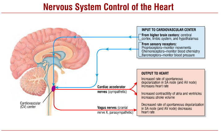
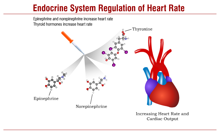
Heart rate is equal to the number of heartbeats per minute. Each heartbeat represents one systole and one diastole, or one cardiac cycle. The autonomic nervous system and the endocrine system regulate heart rate. HR will be calculated from the cardiac cycle length.
Stroke Volume
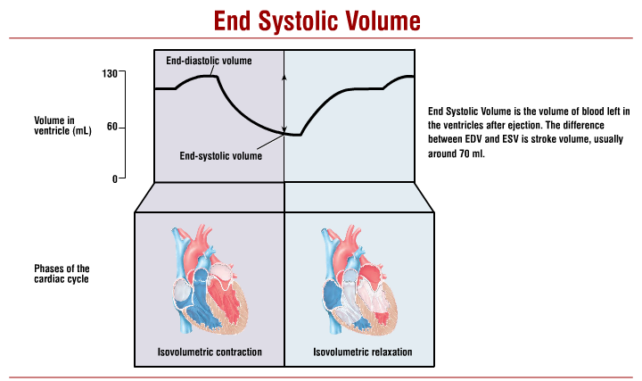
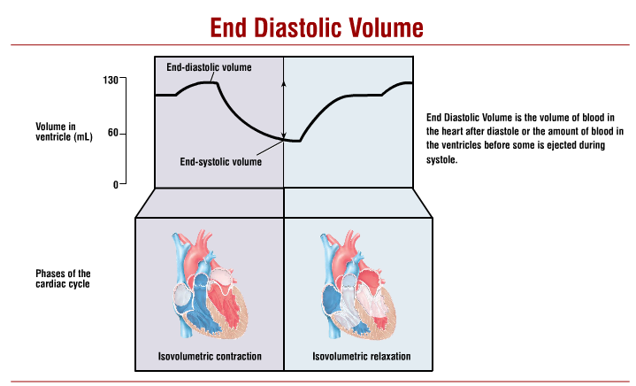
Stroke volume is the amount of blood ejected by one ventricle during one contraction. The stroke volume can be calculated by subtracting the amount of blood remaining in the ventricle at the end of systole (end systolic volume or ESV) from the amount of blood in the ventricle at the end of diastole (end diastolic volume or EDV).
Measuring Cardiac Cycle Length and Ventricular Volumes
A single cardiac cycle includes all the events associated with one heartbeat. Thus, a cardiac cycle consists of systole and diastole of the atria plus systole and diastole of the ventricles.
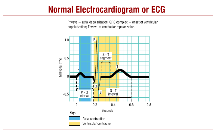
Cardiac cycle length (seconds/heartbeat) will be measured by electrocardiography that produces an electrocardiogram or ECG. Using an ECG, one cardiac cycle is described as being from P wave to P wave. Because this is a cycle and the R wave is the tallest peak, clinicians measure a cardiac cycle from R wave to R wave on an ECG.
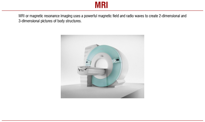
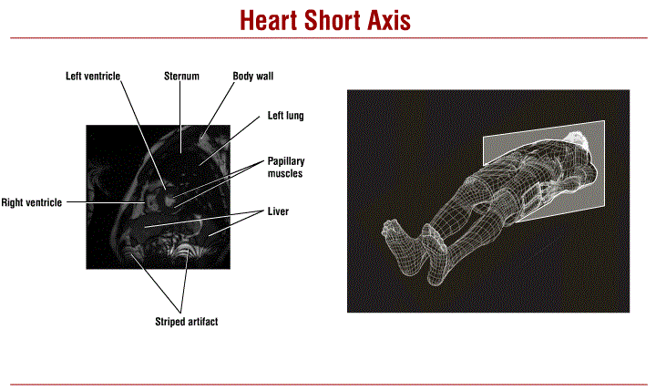
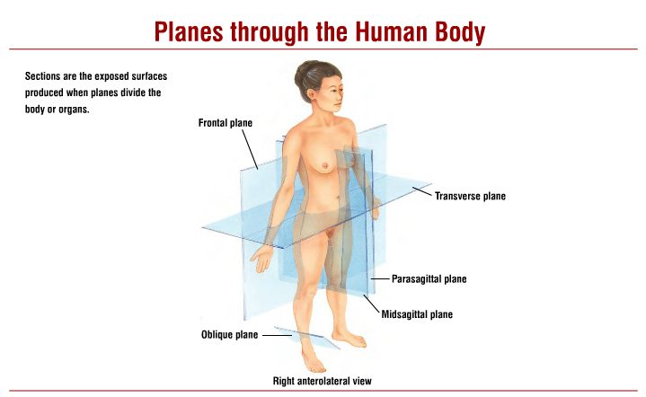
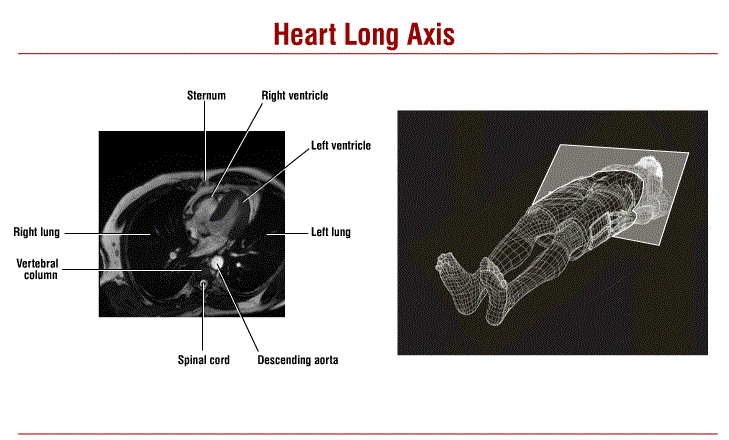
EDV and ESV volumes will be measured from images generated from MRI (magnetic resonance imaging) scans. The MRI produces a series of sectional images through body structures.
The heart short axis MRI view is obtained from a left oblique section through the thorax. The heart long axis MRI view is obtained from a transverse oblique section of the thorax.